Science has relied on dead tissue for a long, long time. The dominant techniques for measuring proteins and molecules require biological tissue to be frozen, fixed, or otherwise destroyed. Researchers slice, stain, probe, and sequence it to read the results.
This practice is extraordinarily powerful, and has shaped our understanding of living systems and advanced countless medical therapies. Yet working with frozen or fixed tissue captures only static snapshots of biology, a moment in time.
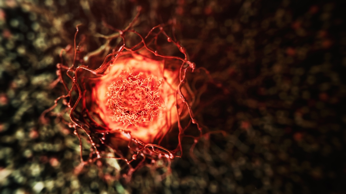
Inflammation – a key driver of deadly diseases, including cancer and heart disease – is not a single moment. It is a dynamic process that begins with subtle molecular signals, unfolds over hours to months, and involves cascading waves of immune activity. When this process becomes dysregulated, it leads to chronic inflammation and tissue damage, processes that remain, for now, invisible to science. Autoimmune and chronic inflammatory diseases are notoriously difficult to diagnose and treat in part because researchers have never been able to observe them unfold in molecular detail in real time.
“This is one of the most significant gaps in the field. Spatiotemporal omics – the ability to sample biomolecules at different locations and times – has taken off over the last few years, but has remained incompatible with live tissue. Existing technologies allow us to look at snapshots, but never the full process.”
Shana Kelley, president of Bioengineering and head of Biohub, Chicago
There is a reason for this gap: The challenge of studying living tissue is genuine. Unlike a frozen sample, live tissue requires oxygen and nutrients and has minimal tolerance for intrusions. Probe a piece of tissue or cell too hard, and one changes the very thing being measured.
So when Biohub put out a funding call to develop technologies for spatiotemporal omics in living tissue, many scientists wrote back to say it was impossible.
Kelley took that as a good sign. At Biohub, high-risk/high-reward science isn’t a liability; it’s the whole idea. Through funding opportunities like this and at Biohub research centers in San Francisco, Chicago, and New York City, scientists are explicitly encouraged to pursue difficult, rate-limiting challenges in biology in order to have the greatest impact on human health and disease.
Drawn from bioengineering, chemistry, immunology, and computation, 15 teams were chosen for proposing compelling, novel platforms that address key technical barriers to spatiotemporal omics, and demonstrate relevance to inflammation, immune regulation, and autoimmune disease models. These teams are now developing a new generation of technologies for minimally invasive, real-time molecular profiling of living tissues. The cutting-edge tools will measure not just where proteins and metabolites are, but when they appear, how they shift, and what their dynamics reveal about inflammation and disease.
The funded projects are strikingly diverse in approach. At the Broad Institute, Paul Blainey and colleagues are engineering T cells inside human tissue explants to continuously report their own molecular activity – a kind of self-narrating biology.
Brent Stockwell at Columbia University is building a platform for real-time lipidomic imaging of inflamed skin, creating a framework for observing, modeling, and reprogramming metabolic pathways and inflammation.
At the University of Oxford, Molly Stevens is developing a real-time, live-tissue analysis platform that layers Raman imaging – an approach for label-free chemical imaging – with novel materials and AI-driven analysis to simultaneously survey the proteome and metabolome of living tissue.
Collectively, these 15 projects form an interconnected portfolio, designed so that a nanoneedle sampling platform developed in London can inform the development of a microneedle proteomic sensor in Boston, and a DNA molecular tomography technique in Chicago can be validated using computational tools built across the network. The goal isn’t just new instruments. It’s a whole new way to measure the immune system in motion.
“We want to watch inflammation in real time – the entire proteome, the entire metabolome, in living tissue,” says Kelley. “It’s white space in the field.”
The 15 teams are:
PI: Amin Ardestani (University of Hull)
Co-PIs: Mohammad Lotfollahi (Wellcome Sanger Institute), Amirpasha Moetazedian (University of Hull)
Instrumented human pancreas: real-time map of autoimmune T1D
PI: Paul Blainey (Broad Institute of MIT and Harvard)
Co-PI: Nir Hacohen (Broad Institute of MIT and Harvard)
Dynamic ex vivo human immunological proteomics
PI: Edward Boyden (Massachusetts Institute of Technology)
Co-PIs: Mriganka Sur (Massachusetts Institute of Technology), Myriam Heiman (Massachusetts Institute of Technology)
Multiplex imaging of astrocytic immune dysfunction in aging
PI: Ciro Chiappini (King’s College London)
Co-PIs: Andy Tay (National University of Singapore), Bart Deplancke (Ecole Polytechnique Federale de Lausanne)
Nanoneedle sampling of live tissue for spatiotemporal omics
PI: Ahmet Coskun (Georgia Institute of Technology)
Co-PIs: Andrei Fedorov (Georgia Tech Research Corp), Khalid Salaita (Emory University)
Spatial mechanomics with dynamic sampling
PI: Liangcai Gu (University of Washington)
Co-PIs: Albert Folch (University of Washington), Zhicheng Ji (Duke University)
Polony gels for submicron spatiotemporal proteomics
PI: Yuhan Lee (Brigham and Women’s Hospital)
Co-PIs: Ang Cui (Brigham and Women’s Hospital), Jeffrey Karp (Brigham and Women’s Hospital)
4D microneedle proteomics for live skin monitoring
PI: Jennifer Rosenbluth (University of California, San Francisco)
Co-PIs: Amrita Basu (University of California, San Francisco), Amy Herr (University of California, Berkeley)
Integrated organoid systems for immunotherapy toxicity
PI: Alex Shalek (Broad Institute of MIT and Harvard)
Co-PI: Fei Chen (Broad Institute of MIT and Harvard)
Nanopillar arrays for live immune signaling capture
PI: Molly Stevens (University of Oxford)
Co-PIs: Jonathan Wojciechowski (University of Oxford), Mark Coles (University of Oxford)
STAMP: Spatial and temporal analysis of the metabolome and proteome
PI: Brent Stockwell (Columbia University)
Co-PIs: Aimee Payne (Columbia University Medical Center), Samuel Sia (Columbia University)
Spatial real-time lipidomic imaging of inflammation in skin
PI: Savas Tay (University of Chicago)
Spatio-temporal proteomics in live immune tissues
PI: Sihong Wang (University of Chicago)
Co-PI: Cathryn Nagler (University of Chicago)
HydroSense: A hydrogel bioelectronic platform for in vivo omics
PI: Joshua Weinstein (University of Chicago)
DNA-based approaches for molecular mapping in live tissue
PI: Yi Zhang (University of Connecticut)
Co-PIs: Robert Kennedy (University of Michigan), Sasan Jalili (Jackson Laboratory)
3D push-pull microsampling networks for spatiotemporal omics