Dynamic Structural Cell Biology
Our group aims to generate deep, mechanistic insights into complex biological systems by resolving how molecular interactions give rise to cellular function.
We are inventing, optimizing, and deploying next-generation imaging technologies that enable interrogation of cellular structure in three dimensions at near-atomic resolution. These technologies will be made robust, scalable, and accessible, enabling structural cell biology at global scale.
Research
Goal 1: Enable the Structure of the Cell to be Determined at Atomic Resolution
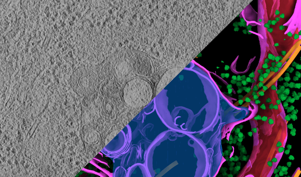
Cryo-electron tomography (cryo-ET) can reveal molecular structures inside cells, but today it is slow, highly specialized, and not up to the task. We are developing advances in imaging, sample preparation, distortion correction, and AI analysis to enable real-time results, dramatically increase throughput, and make cryo-ET a scalable, accessible tool for cell biology.
Optimize sample preparation and labeling
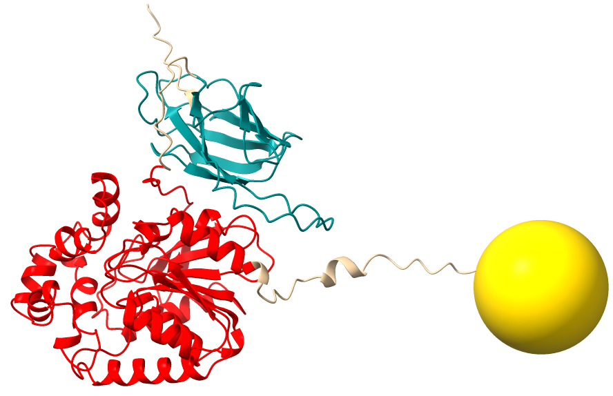
Current cryo-ET methods can visualize only the largest cellular complexes, representing a tiny fraction of the cellular proteome. Yet much of cell biology derives from smaller and low copy number complexes associated with specific cellular regions. Finding these requires the development of high-affinity labels. We are developing new labeling, sample preparation, and cryo-CLEM tools.
Optimize targeting of cellular structures
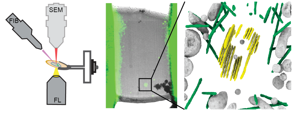
We are building AI- and CLEM-guided targeting and analysis tools to precisely locate, image, and reconstruct rare molecular structures in cryo-ET samples.
Improve image contrast via dual laser phase plate (xLPP) and new cameras for molecular clarity
We have built a dual laser phase plate (xLPP) that will greatly enhance image contrast in cryo-electron microscopy (cryo-EM) without compromising high-resolution information. Read the preprintand the press release.
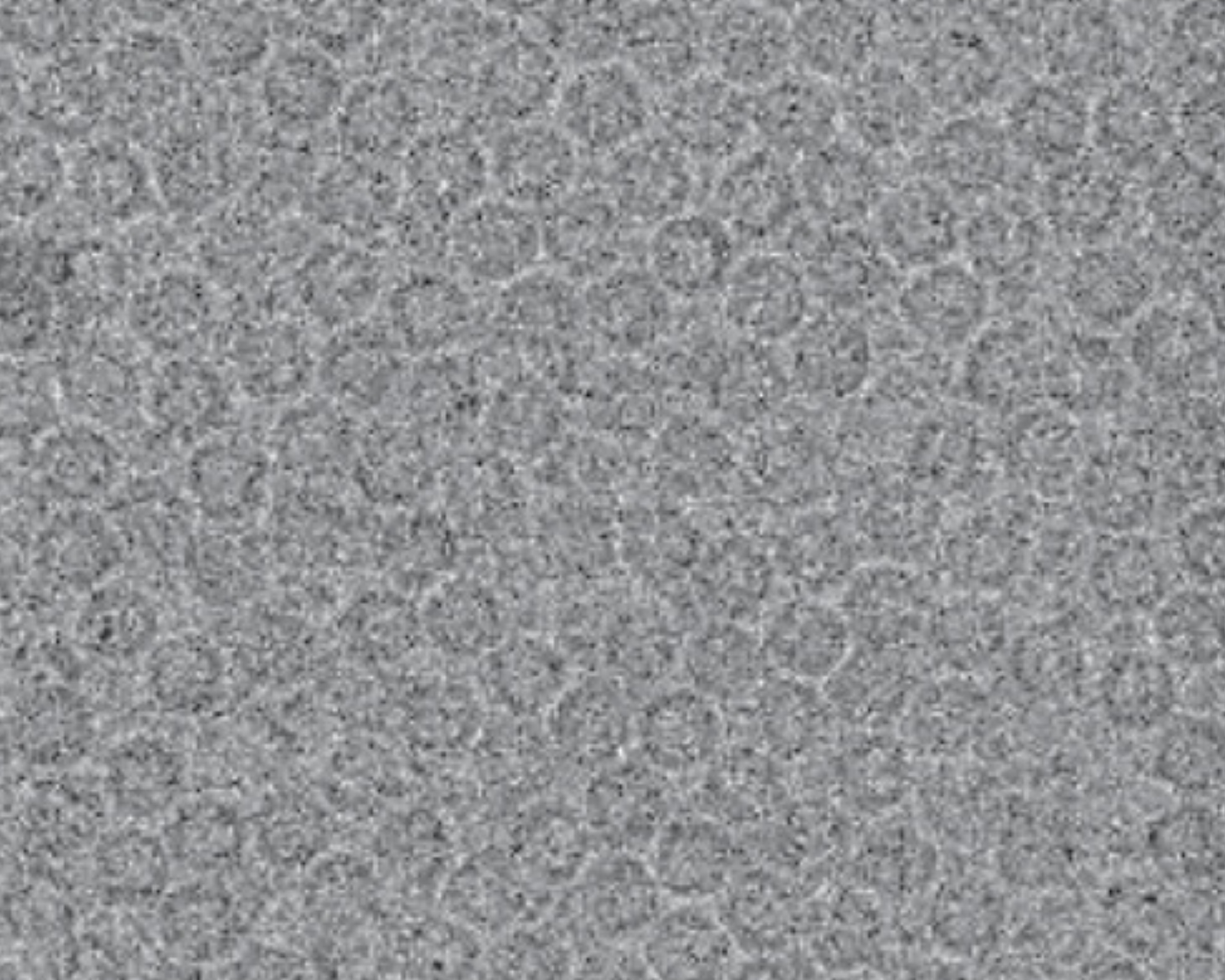
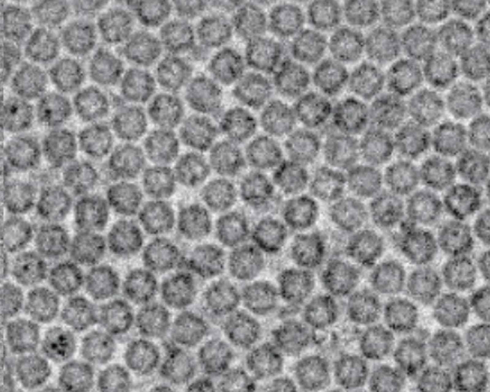
Major gains in image information content and protein detectability can be achieved via a routinely usable laser phase plate, a brilliant invention by Holger Müller and his team at UC Berkeley. While proof-of-concept has been well demonstrated, significant optimization of the device, the microscope, and the control systems are required to make this exciting technology routinely usable for cryo-ET. In addition, new cameras promise to significantly enhance data quality and collection efficiency.
Develop optimized pipelines with a unified web-based interface to accelerate cryo-ET throughput
Cryo-ET workflows are often complex and fragmented across many tools. We are streamlining the entire process within Embrella, a unified web platform that integrates data collection, processing, annotation, analysis, and interactive visualization.
Improve raw tomogram resolution through advanced denoising and deformation correction
We are improving cryo-ET image processing to dramatically increase tomogram resolution. By combining enhanced image contrast from the xLPP (dual laser phase plate) with advanced denoising and motion-correction algorithms, we aim to improve raw tomogram resolution from 30–40 Å to 15–20 Å, enabling more accurate visualization, segmentation, and structural analysis.
Develop AI-guided tools for template matching, segmentation, and object finding
We are building AI tools that combine structural predictions, imaging data, and labeling information to accurately detect and classify molecular complexes in challenging cryo-ET datasets.
AI tools are being reframed to:
- Produce standardized structural descriptors suitable for archetype-based modeling.
- Use PPI- and OpenCell-informed priors to guide object detection.
Implement a holistic AI framework to simultaneously detect and refine structures
By leveraging foundation AI, we seek to unify molecular identification and structure determination into a single process, transforming how cellular structures are discovered and analyzed in situ.
This framework is now being explicitly defined as the junction between:
- Structural discovery (cryo-ET)
- Interaction networks (MS-based PPI, proximity labeling)
- Model building via guiding foundational protein models
- Archetype representations of cell state
Integrate and deliver validated data and annotations on a standardized data portal
We have created an open-access cryo-ET data platform with standardized datasets, rich annotations, and interoperable metadata to accelerate biological discovery and machine learning across the community.
The portal is positioned to:
- Provide high-quality training data for segmentation, object detection, and virtual cell modeling
- Enable cross-institutional annotation aggregation to expand ML training set diversity
- Link structural annotations to PPI networks through standardized ontology terms
- Connect to complementary resources (e.g., Proteohub) as multimodal integration matures
Goal 2: Bridge the Resolution Gap Between Light and Electron Microscopy and Visualize Cellular and Tissue Context
By bridging cryo-ET and light microscopy, we aim to link molecular structures to cellular and tissue context, creating a seamless view across biological scales.
Cryo blockface imaging with improved protein/DNA contrast
By advancing cryo-SEM contrast and detection technologies, we aim to visualize all major cellular components across whole cells and tissues at nanometer-scale resolution, providing a powerful bridge between molecular and cellular imaging.
Atomic force microscopy (AFM) readout for novel contrast modes (topography, polarizability)
By integrating AFM with cryo block-face imaging, we aim to unlock new sources of biological contrast, providing richer structural information across cells and tissues at the nanoscale.
Near-field optical readouts for chemical/protein of interest (POI) information
Integrating tip-scanning optical methods with cryo block-face imaging could reveal both structure and chemistry, enabling super-resolution molecular mapping and new forms of biological contrast across cells and tissues.
Tools
We make our biological imaging tools and datasets freely available to the community.
AreTomo2
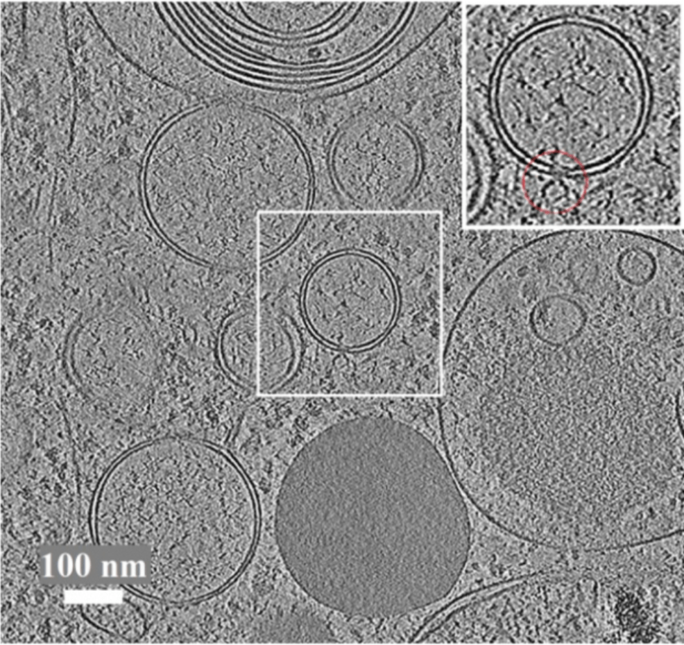
AreTomo2, a multi-GPU accelerated software package, enables full automation of tomographic alignment, reconstruction, and robust per-tilt CTF estimation. These functions are fully integrated in a single software package. AreTomo2 strives to be fast and accurate. Its high throughput can be used for on-the-fly reconstruction, providing researchers with a new opportunity to assess and increase their sample quality in real-time.
AreTomoLive
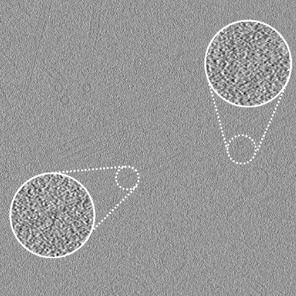
AreTomoLive is an automated real-time pipeline to advance cryo-electron tomography and subtomogram averaging. It is composed of two GPU-accelerated packages: AreTomo3 streamlines tomographic alignment and reconstruction and DenoisET performs contrast enhancement.
Copick
copick is an open-source Python framework for managing and annotating cryo-electron tomography data at scale. It offers a unified interface to tomograms, segmentations, picks, and meshes across local and cloud storage, with integrations for tools like ChimeraX and napari to support reproducible annotation workflows.
CryoET Data Portal
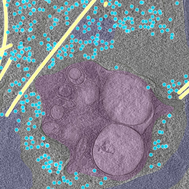
The CryoET Data Portal is a cloud-based, open-source portal aimed at driving the development of automated annotations of cryoET datasets. This tool has the potential to shorten data processing time from months or years to weeks.
GCtfFind

GCtfFind is a GPU-accelerated software package for robust estimation of the contrast transfer function (CTF) of cryo-ET tilt series and cryo-EM micrographs, essential information needed for cryo-ET subtomogram averaging and cryo-EM single-particle reconstruction.
MotionCor3
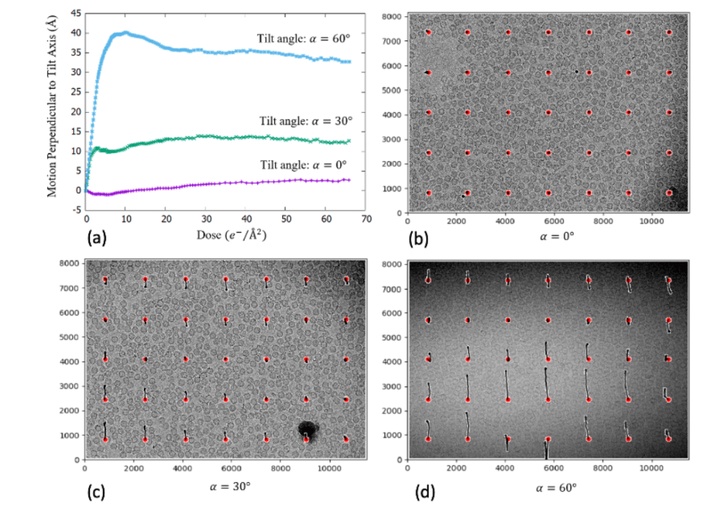
MotionCor3 is a multi-GPU accelerated program that corrects anisotropic beam-induced sample motion at the single pixel level, suitable for both single particle and tomographic images. MotionCor3 enables motion-corrected images that keep pace with data collection.
OCTOPI
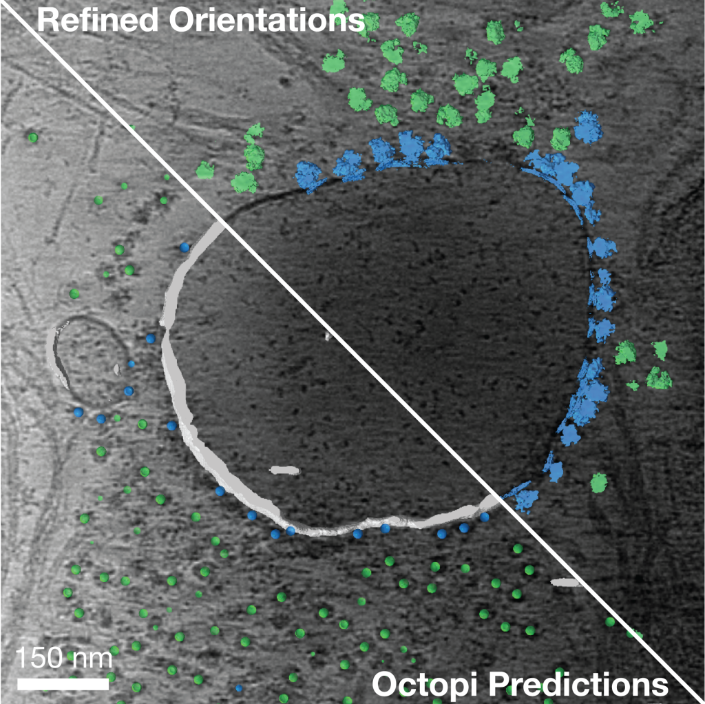
OCTOPI is a platform for training supervised 3D convolutional neural networks for volumetric annotation and particle picking. Built to work seamlessly with labeled data (e.g., generated via SABER or nnInteractive), OCTOPI includes a self-supervised framework that helps identify optimal network architectures for a given dataset, enabling efficient and high-quality model development even with limited annotations.
SABER
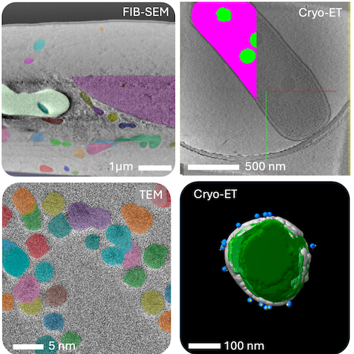
SABER is a flexible segmentation framework that enables rapid 2D and 3D annotation with minimal training effort. It leverages precomputed segmentations from state-of-the-art “Segment Anything” models (e.g., SAM2/SAM3) and allows users to efficiently assign semantic labels to objects of interest. This dramatically accelerates the creation of labeled datasets for downstream analysis.