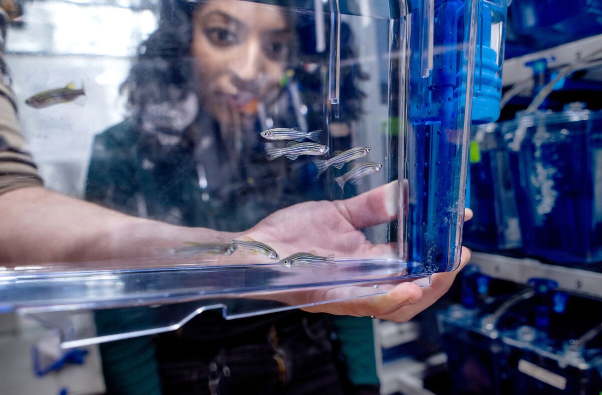
Royer Group: Organismal Architecture
How do organisms develop from a single cell into a fully functional body, with billions of self-organizing cells that form tissues and have different functions?
Our Research
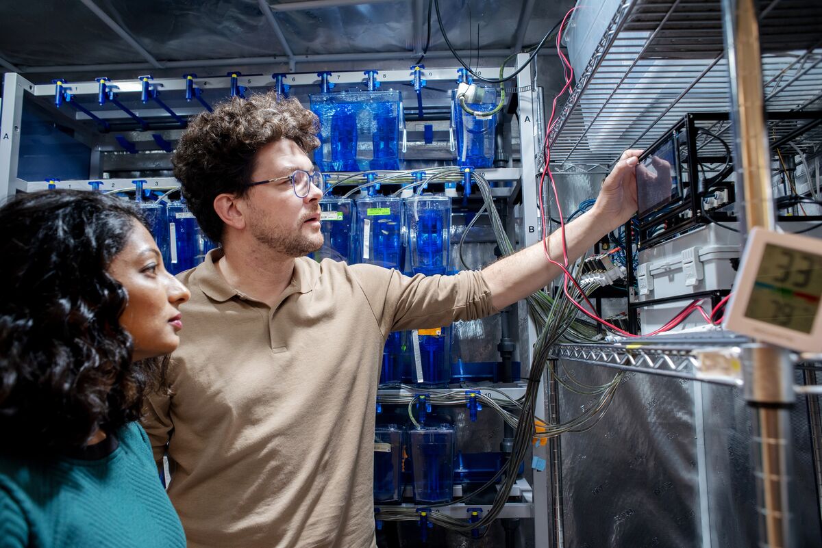
We are a pluridisciplinary team of computer scientists, optical engineers, and biologists.
Our goal is to build a time-resolved, multidimensional atlas of vertebrate development using zebrafish as a model organism.
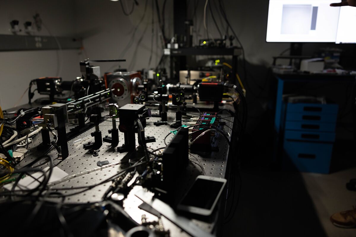
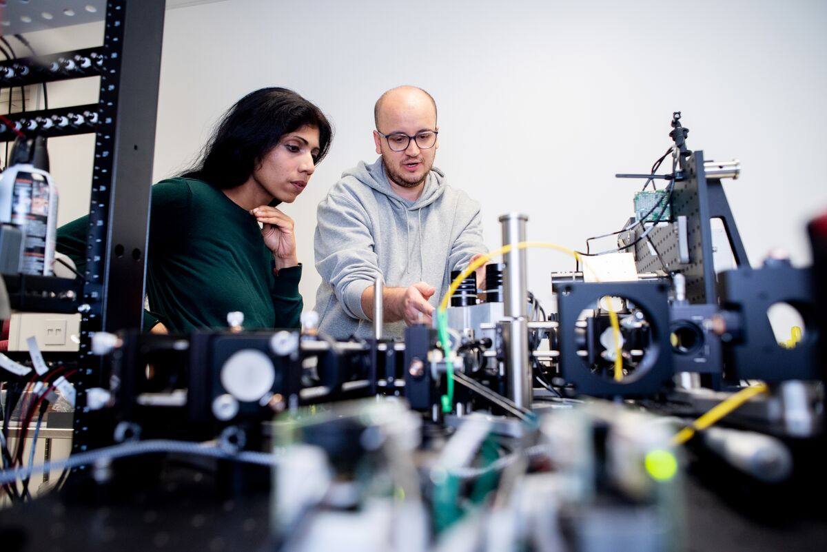
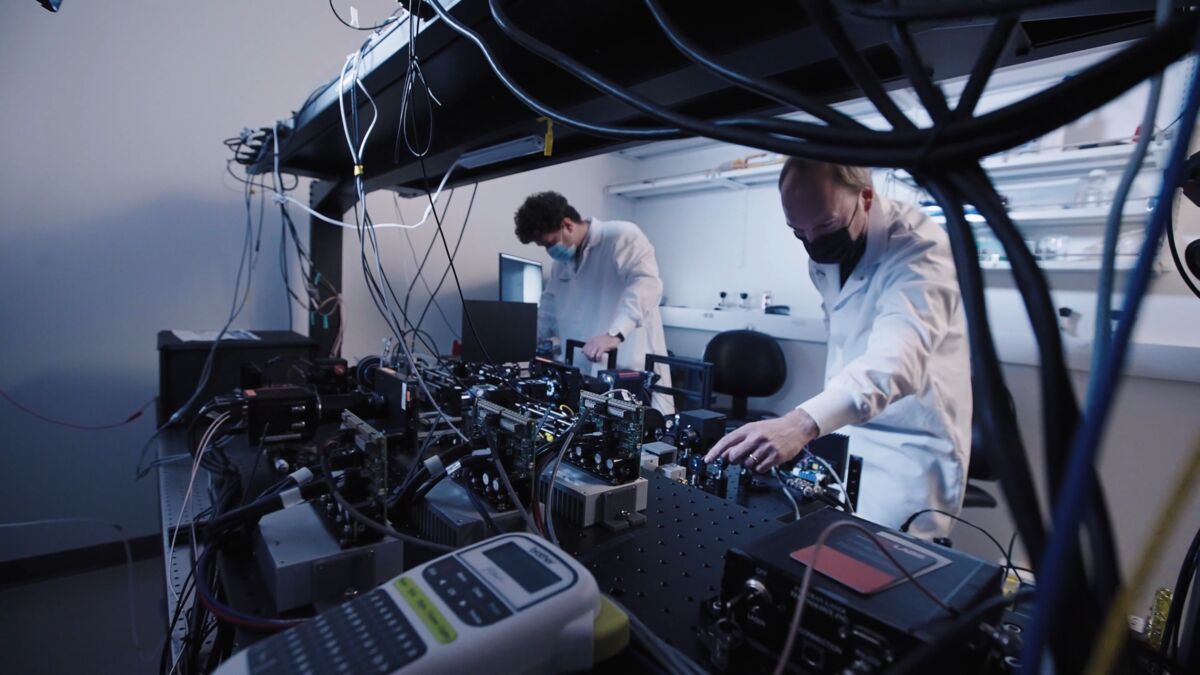
To attain this goal, we design, build, and implement novel state-of-the-art light-sheet microscopes, and employ and also design deep learning–based image processing and analysis algorithms.
Bioimage Informatics
Deep learning-based image restoration, 5D bioimage visualization, GPU-accelerated image processing
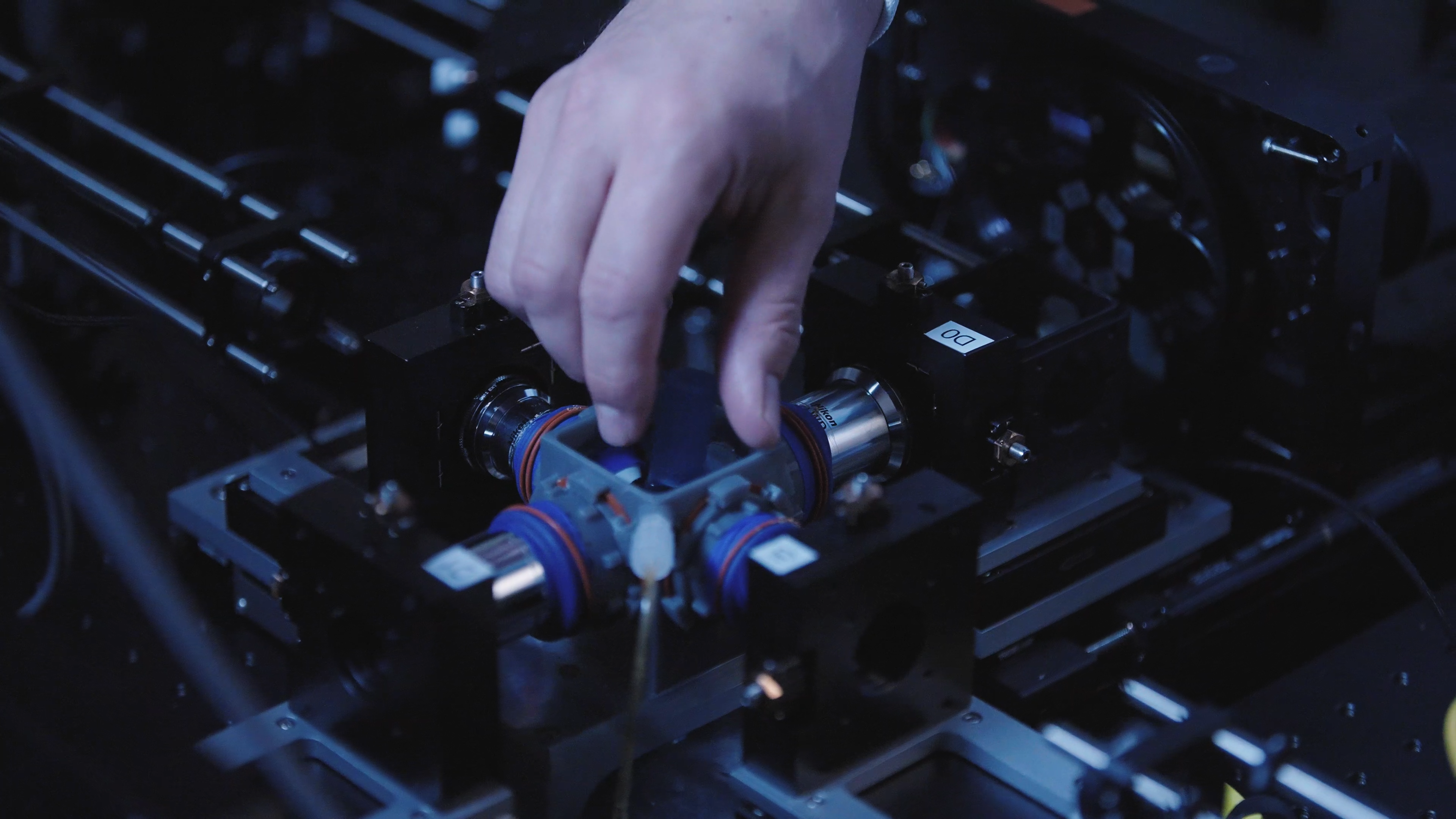
Advanced Microscopy
Light-sheet microscopy, single objective microscopy, adaptive imaging, smart microscopy
Developmental Biology
Cellular atlas of vertebrate development, Zebrafish development
Selected Papers and Projects
Image Restoration

Nature Methods, 2018
Weigert et al.
Nature Methods, 2019
Beltangady and Royer
PMLR, 2019
Batson and Royer
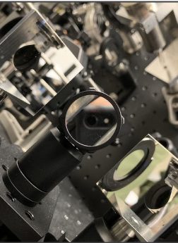
Image Processing & Analysis
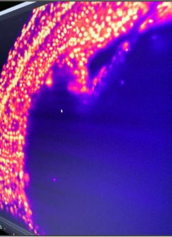
bioRxiv, 2024
Bragantini et al.
Nature Methods, 2022
Kobayashi et al.
Nature Methods, 2019
Haase et al.
napari
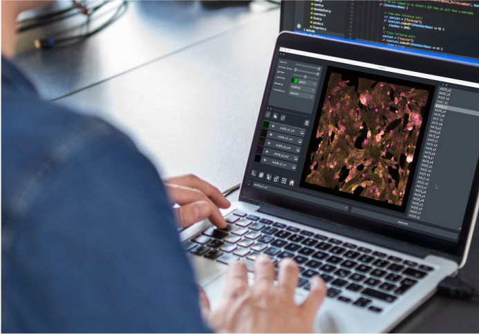
Created by Loïc Royer, Juan Nuñez-Iglesias of Monash University, and Nick Sofroniew of the Chan Zuckerberg Initiative, napari is an open-source collaboration involving Biohub, the CZI Science Technology team, and many other scientists and computational biologists around the world.